Educational scope. This article supports professional learning and case discussion. It is not patient-specific advice, a clinical protocol or a substitute for supervised training.
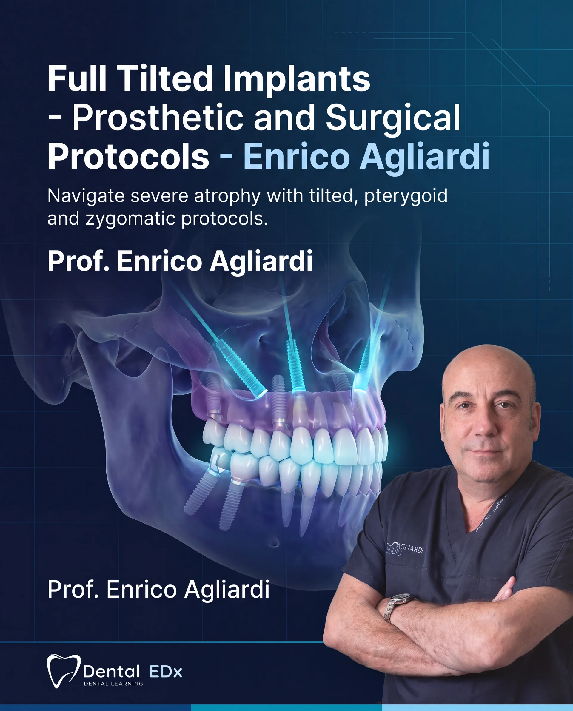
A tilted trajectory is visually distinctive, but the image alone does not explain why it was chosen. Advanced training should connect the proposed trajectory with the complete anatomical and prosthetic problem, including alternative approaches.
CLINICAL TAKEAWAY
What matters most
- Identify the problem being solved.
- Separate different anatomical approaches.
- Look for prosthetic follow-through.
EVIDENCE LENS
The edentulous-maxilla consensus addresses multiple planning dimensions; no single feature of a surgical image explains the entire rehabilitation.
Ask what the trajectory solves
In a case presentation, identify the stated problem before examining the implant angle. Record the alternatives that were considered and the reasons for selecting this approach. Avoid assuming that the same visible trajectory has the same rationale in another patient.
Distinguish related advanced techniques
Tilted, pterygoid and zygomatic approaches should not be collapsed into one interchangeable procedure. Read the lesson descriptions closely and identify which anatomical regions and treatment scenarios each covers. Course breadth does not eliminate the need for technique-specific training.
Assess the restorative explanation
Look beyond placement to the prosthesis and the means of evaluating it. Ask whether the presentation includes complications, changes of plan and later review. For your learning plan, identify the knowledge gaps that need supervised instruction before the topic could become part of your clinical scope.
Angulation is part of a system, not an isolated trick
A tilted implant may be discussed in relation to anatomy, prosthetic support and avoiding additional procedures. The rationale should be evaluated within the complete arch plan, including connection geometry, restoration design and maintenance.
Look for anatomy-specific escalation and referral
Advanced training should explain the imaging, experience and complication planning required for the approaches it demonstrates. A learner should be able to identify where observation ends and supervised clinical development or referral begins.
Turn the topic into a structured team review
Choose one de-identified case that raises the question of tilted implants. Review it in stages rather than jumping to the final image: define the patient-centered objective, list the available records, identify who made each decision and mark every assumption or missing item. Then compare the sequence with the cited evidence and the applicable manufacturer or professional guidance. The objective is to understand why this pathway was selected, not to reproduce a technique from an article.
Repeat the review after delivery or follow-up and ask what the team would change. Use these checkpoints: Identify the problem being solved. Separate different anatomical approaches. Look for prosthetic follow-through. Record disagreements between the clinical, technical and maintenance perspectives instead of forcing an immediate consensus. A short, documented calibration exercise makes the article useful to the whole team and creates questions that can be taken into formal education, mentoring or supervised practice.
TAKE INTO YOUR NEXT CASE DISCUSSION
Three points to keep in view
- Identify the problem being solved.
- Separate different anatomical approaches.
- Look for prosthetic follow-through.
Evidence & references
The edentulous-maxilla consensus addresses multiple planning dimensions; no single feature of a surgical image explains the entire rehabilitation.
- Consensus on rehabilitation of the edentulous maxilla: number of implants, placement and loading Global Consensus for Clinical Guidelines
References support the evidence discussion, not an endorsement of DentalEDX. Practical study prompts are editorial suggestions. No patient outcomes or professional accreditation are promised.
A common question
Can I judge suitability from a panoramic image alone?
A teaching image is not a substitute for a complete, appropriately interpreted clinical assessment.
CONTINUE LEARNING
Explore this topic in more depth.
Review the curriculum and prerequisites to choose teaching that fits your experience.